The postmortem examination technique, also called necropsy, is not complicated. I like to start by placing the carcass on its left side, cutting the dead animal in the right axilla, and proceeding to open the skin from the mandible to the inguinal region.

It is better to cut the skin from the subcutaneous tissue to maintain the sharpness of the knife edge. After the cadaver is skinned, you open every cavity: abdomen, thorax and pericardium. In every case, I always try to do a complete necropsy to make sure I do not overlook anything.
I try to make sure that the people I train are enthusiastic about the postmortem examination, because it is a very important component of the diagnostic process. For the exam to be complete it is important to use all your senses. You have in front of you the outside and the inside of the animal for you to examine, touch and even smell.
I remember a group of four heifers presented to the postmortem service on a very cold morning. The history was not very specific. Two animals were found dead; the other two presented with difficulty breathing for a couple of days. All of them were depressed and had poor appetite.
The postmortem examination gives you access to all the organs that you need to sample, and this is the moment where you need to think about what tissues or tests should be submitted in order to resolve the case.
When we opened the carcasses of the four heifers, we found that the pathological changes were centered in the thoracic cavity. All animals had some degree of lung disease. How did we know that? The short answer is because I touched and felt the lungs.
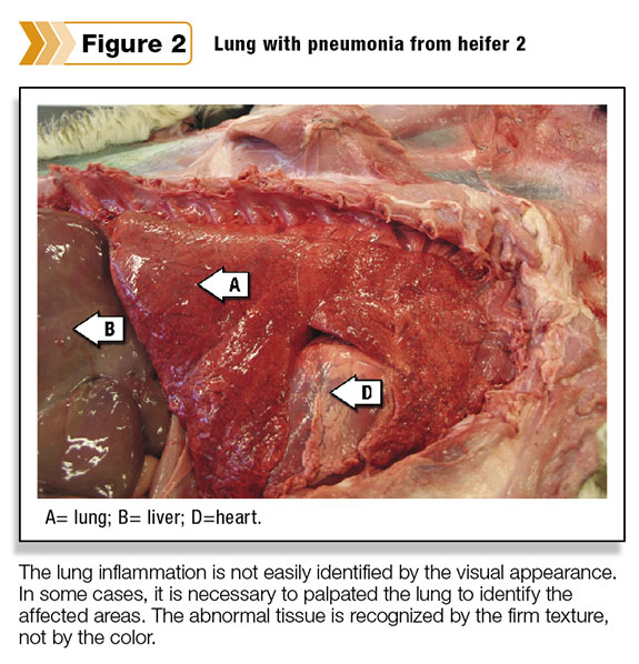
Respiratory disease is challenging because you need to feel the tissue. You should not be afraid to touch the lungs. The lung might not look affected to the naked eye, but it will feel affected if there is pathology. In order to properly assess some lesions you need to develop some scale of tissue texture and resistance.
The guidelines I use are: Soft is your lips (which is primarily muscle), firm is your nose (which has more cartilage) and hard is your forehead (which is bone). I always remind my trainees to touch those places before the postmortem examination (not during the examination).
If you remember the different consistencies that you can find during the postmortem examination, you can incorporate that information in your interpretation of the findings. When it comes to lung disease, one of my best friends says: “If it ain’t firm, it ain’t pneumonia.” That is because the lung with pneumonia feels firm to the touch, as firm as when you touch your nose.
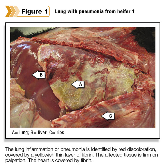
In our case, the four animals had firm areas mainly in the anteroventral portion of the lung. Not all of the lungs looked affected, but all of them felt firm ( Figures 1 and 2 ). The most common reason for lung disease or pneumonia is bacterial infection.
I believe that when heifers are exposed to a drastic drop in temperature it is difficult for them to adapt and it will be easier for the bacteria to invade via the airways.
In these cases, the area of the lung that is most likely affected is the anteroventral part. Some books describe this type of lung disease as enzootic pneumonia.
We diagnosed lung disease in three of the four
heifers. In addition, one of the heifers showed a superficial thin layer of yellowish tissue over the lungs, which is fibrin. Fibrin is the same protein that will help to clot your blood when you cut yourself.
The abnormal lungs were always firm in the affected areas because the tissue is filled with inflammatory cells and proteins within the airways, where they are not found in healthy animals.
The affected lung is also more friable to manipulation, and it will not float when a little piece is dropped in a container with some water.
To complete the diagnosis, lung tissue was submitted for culture to identify the potential organism involved. In our case, we isolated a bacterial organism called Pasteurella, a common pathogen in cow lungs.
A postmortem examination might sound like an unattractive procedure, but if you know what you are doing, it can be very rewarding. Plus it does not require fancy equipment – only one knife is needed. It is a simple technique and one that has not changed much since the first necropsies were performed.
Necropsy is a simple way to aid in disease diagnosis. It allows you to collect information to corroborate the clinical presentation. The information comes from the gross or macroscopic appearance of the different tissues and organs inside the body. It is like detective work, where you dissect in detail all the cavities and at the end you try to put the pieces of the puzzle together.
Conducting a postmortem examination of our four heifers allowed us to understand why the animals died or were depressed or had difficulty breathing. In our case, the findings were centered in the lung, and the consistency of the tissue allowed us to describe the pathological changes. We use a lot of common sense, plus our five senses, when doing postmortem exams. In a future article, I will tell you about a case in which the sense of smell proved very useful.


