Mastitis is an inflammatory disease of the mammary gland that impacts all food production animals reared for milk harvest, but is well documented as the most widespread and costly animal health problem in dairy cows.

Improved sophistication of mastitis detection tools has made great strides in the control of contagious pathogens and knowledge of environmental pathogens. One such tool is culturing and the use of a milk quality laboratory. Microbiological culture has been the gold standard for many years, but diagnostic methods have evolved over time, as have the pathogens themselves. So what does it all mean for the producer and their herd health veterinarians, making daily management decisions?
The primary goal for most producers of using a milk quality laboratory service is to create data-driven management decisions. Traditional microbiological culture of milk samples remains the most common and economic diagnostic for mastitis, but molecular methods such as PCR or mass spectrometry (Maldi-ToF) are becoming increasingly widespread.
The clear advantage of using molecular detection is rapid turnaround time and speciation, but the applicability of some results generated by molecular methods may not be practical for making daily management decisions. For example, speciation results generated by Maldi-ToF are useful in specific bacterial investigations but do not necessarily translate into cow-level management decisions. Conversely, the data being generated by these methods is changing our epidemiological understanding of pathogens such as Mycoplasma, Staphylococcus aureus and Prototheca, which plays a very important role in how they are managed.
Staphylococcus aureus can be easily detected via traditional culture methods within 24 to 48 hours of taking the sample and has traditionally been identified by the classic “beta” hemolytic zone surrounding the colony; however, there are absolutely strains which do not express this characteristic. Since beta-hemolytic S. aureus has historically been the dominant field strain, some laboratories may not be routinely using secondary coagulase testing to find these alternative strains as a part of their detection protocol.
Analysis of results generated over a five-year period from routine samples brought into Udder Health Systems laboratories reviewed almost 1 million cow sample records to take a closer look at the prevalence and detection between different strains. A total of 23,367 were confirmed as S. aureus-positive by either colony morphology, coagulase testing or by use of Maldi-ToF. Of those S. aureus-positive colonies, only 48% were identified by seeing the classic beta-hemolytic zone, whereas 52% of the positives did not. Enhancement was used on all samples with no growth at 24 hours, and 18% of all S. aureus colonies found were a result of enhanced samples.
The discovery that greater than 50% of S. aureus colonies were not beta-hemolytic was a surprising and biologically significant observation. This suggests laboratories not routinely coagulase testing are missing detection of a substantial number of S. aureus-infected animals, which are potentially spreading within the herd and causing milk quality degradation.
Like control of S. aureus, traditional culture for Mycoplasma has served the industry well, but the increasing use of molecular technology is changing the epidemiological picture. Control of Mycoplasma mastitis has historically been challenged by slow growth (seven- to 10-day incubation) on culture, false-positive contamination with Acholeplasma spp. and intermittent shedding patterns.
Acholeplasma is a recognized environmental contaminant which may end up in the milk sample because of poor sampling technique, and it is indeterminate if this organism ever results in mastitis cases. Acholeplasma grows under the same conditions as Mycoplasma, and it cannot be differentiated from pathogenic species with the naked eye; confirmation testing must be done by PCR (rapid) or digitonin culture test (seven to 10 days).
The use of PCR allows detection and speciation of the pathogenic strains of Mycoplasma (M. bovis, M. bovigenitalium, M. alkalescens, M. californicum, M. canadense and M. arginini) and real-time differentiation from Acholeplasma, within hours of receiving a sample. Analysis of data from more than 6,000 colonies identified as culture-positive from individual cow samples submitted for routine screening to the Udder Health Systems laboratories found that approximately 42% were Acholeplasma spp. In this same dataset, 1,216 colonies from culture-positive bulk tank milks were also tested, but less than 2% were found to be Acholeplasma spp. (Table 1).
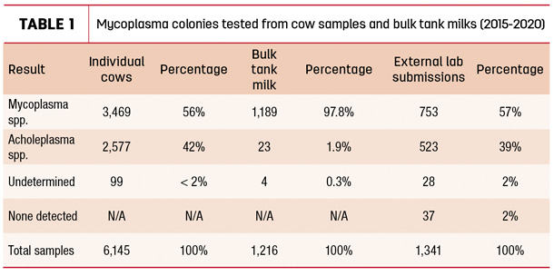
Another dataset comprising of 1,341 colonies submitted for speciation from external laboratories had similar findings when analyzed; 39% were Acholeplasma spp.
There are numerous laboratories that offer a confirmation service on bacterial isolates submitted to them, who may be of assistance to laboratories which do not have PCR capacity. Alternatively, the digitonin test is a time-consuming but inexpensive culture-based method for differentiating between the two species that would be feasible for most labs. Laboratories not routinely doing confirmations of Mycoplasma colonies may be reporting false positives and inadvertently sending healthy animals to slaughter.
Prototheca mastitis has received an increasing amount of attention in recent years as a causative agent of both clinical and subclinical mastitis. Prototheca colonies are easily missed on standard blood agar culture due to slow growth and cows often being low shedders, or they may be misidentified as Staphylococci colonies.
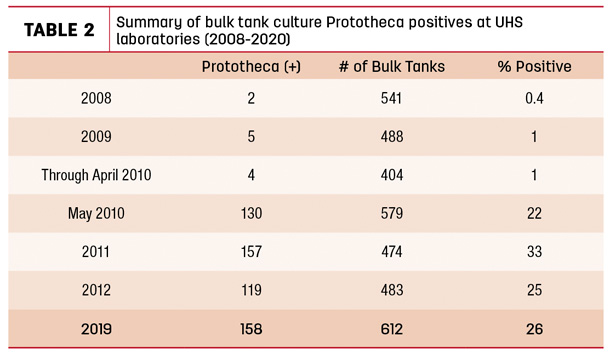
Use of a selective Prototheca agar will significantly enhance a laboratory’s ability to detect Prototheca at both the bulk tank and individual cow level. In 2010, our lab incorporated a selective Prototheca agar into routine bulk tank culture screening. Before that, the observed prevalence of bulk tank cultures positive for Prototheca was less than 1%. Following that change to procedure, observed prevalence increased to 22% to 25% and has remained at that level across all bulk tank results from our laboratories (Table 2).
The significant increase in Prototheca detection at the bulk tank level spurred further investigation at the individual cow level. A review of nearly 200,000 cow samples cultured on only blood agar between 2015-20 in our labs identified 251 Prototheca isolates, which is a very low 0.14% prevalence. During the same time period, an additional 106,000 cow samples were cultured on selective Prototheca agar and, of these, a total of 2,676 isolates were identified, for a prevalence of 2.5%.
By comparison to other contagious pathogens, the overall prevalence remains low. However, the increase in detection by adding a selective screening method should be noted. Environmental areas of importance include water, manure and mud in alleys and lounging areas but, additionally, two critically important sources of exposure are bedding material and infected animals themselves. Routine bedding cultures done in most laboratories will not find Prototheca, which has been the experience in our laboratories as well. This has been changed by the incorporation of enhancement methods and selective agar.
Over a two-year period of using specific Prototheca detection protocols on water, bedding and other environmental samples, Prototheca was detected in 40% of submitted samples. In particular, sand and recycled sand bedding samples were problematic sources found to be positive and a likely source of new infections. In contradiction to historical belief, it was rarely detected in water samples. It should be noted that detection of Prototheca at the bulk tank level is responsible for specific investigations into cows and environmental samples and not part of routine screening.
The use of a laboratory service is an essential tool for progressive dairies to achieve the milk quality standards and goals they set for themselves. For dairies both large and small, laboratory results from individual cow, bulk tank and environmental cultures will allow them to make smart and economical management decisions. However, mastitis pathogens and the way we find them does not look the same as it did 50 years ago.
As milk quality professionals and consultants, we need to be evaluating if we are asking the right questions, both in conversation and in our detection methods. Veterinary and milk quality laboratories have a responsibility to deliver accurate and quality data that will serve the needs of both dairy managers and the health of their animals.
References omitted but are available upon request by sending an email to the editor.
Take-home messages
- Laboratories not routinely coagulase testing are missing detection of a substantial number of S. aureus-infected animals, which are potentially spreading within the herd and causing milk quality degradation.
- Laboratories that are not routinely doing confirmations of Mycoplasma colonies may be reporting false-positives and inadvertently sending healthy animals to slaughter.
- Selective screening for Prototheca may be warranted. Environmental areas of importance include water, manure and mud in alleys and lounging areas – but additionally, two critically important sources of exposure are bedding material and infected animals themselves.


