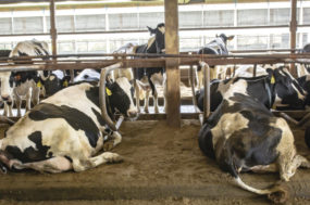
Mastitis is an inflammation of the mammary gland or udder tissue. Inflammation is the response of the body to injury. In cows, this response (i.e., mastitis) is usually provoked by infection with bacteria. Mastitis can also be the result of noninfectious causes, such as mechanical damage. A poorly adjusted milking machine or narrow stalls and poorly trimmed claws may cause mechanical injuries to the teats and the udder.
Even when bacteria are not present, the body will respond to such injuries with inflammation, much like the redness, swelling and pain associated with a twisted ankle. Mechanical damage and bacterial infections can both result in mastitis. Furthermore, mechanical damage may open the way for bacterial infections.
The response of the body, the inflammation, may be visible or invisible. If a response is visible, the mastitis is considered to be clinical. Clinical signs can be mild, moderate or severe. In mild cases, visible abnormalities are limited to the milk only (e.g., clots, flakes or watery milk).
Of course, one has to look to see such changes. If cows are not forestripped before the milking unit is attached, mild clinical mastitis will go unnoticed. In the case of moderate clinical mastitis, both milk and udder show abnormalities. The udder may show redness, swelling or pain and can be warm to the touch. Usually, the function (milk production) is affected, too.
Redness, swelling, pain, increased temperature and abnormal function are the typical characteristics of inflammation. In severe cases milk, udder and cow are affected. The animal may have a fever, be off-feed, depressed and down. Very severe cases are also described as toxic cases.
Some herds only consider the severe cases to be clinical cases, thus underestimating the real number of clinical cases. When discussing or comparing the number of clinical cases, make sure everybody has the same understanding of what they mean by clinical mastitis.
Severe clinical mastitis is often called acute mastitis. Strictly speaking, acute refers to the duration of the mastitis, rather than to the severity. Clinical mastitis can be acute (just started) or chronic (e.g., chronic E. coli mastitis).
When no signs of clinical mastitis are visible, the mastitis is subclinical. Because subclinical mastitis often goes unnoticed and unaddressed, the duration of subclinical mastitis is usually longer than the duration of clinical mastitis. For many people chronic mastitis and subclinical mastitis are more or less synonymous, although the first term refers to duration and the latter term to severity.
Even though subclinical mastitis can’t been seen with the naked eye, it is very important. It is associated with high somatic cell count (SCC) and production losses, and subclinical mastitis cases can be a source of infection for other animals in the herd.
Many subclinical mastitis cases show up as clinical mastitis cases when a cow is in heat, when there is a change in the weather or at some other point in time. High SCC are associated with lower milk and cheese yields, shorter shelf life of processed milk and lower milk prices. Usually, 200,000 cells per milliliter is used as a threshold between normal and abnormal milk, but any cow level SCC over 50,000 cells per milliliter is associated with a decrease in production relative to the cow’s genetic potential.
In practical terms, milking cows with high SCC means you are milking more cows than necessary to produce the same amount of milk. Each extra cow needs to be housed, managed, fed, inseminated, vaccinated and milked.
From an economic point of view, subclinical mastitis is probably more important than clinical mastitis, even though we cannot see it in the milking parlor. To see subclinical mastitis, additional tests such as SCC measurement, CMT (California Mastitis Test or paddle test), culture of bacteria or conductivity measurements must be used.
For mastitis management, the way mastitis spreads is more important than the way it shows up. Once we understand how and where cows become infected with bacteria, we can take management action to prevent infections and new mastitis cases from occurring.
Bacterial mastitis is characterized in two groups, based on the way the bacteria are spread. On the one hand, there is contagious mastitis. For contagious mastitis, the cow’s udder is the main source of bacteria. Spread of bacteria mostly happens during milking, via udder cloths, liners or milkers’ hands.
To solve a contagious mastitis problem, the source of bacteria needs to be identified and removed. In other words, infected cows are detected based on high SCC and culture results, and these animals are subsequently treated, culled or segregated from the rest of the herd.
On the other hand, there is environmental mastitis. As the name implies, the environment is the main source of bacteria in this scenario. The environment includes the cow itself: manure, skin, mucous membranes and anything outside of the udder, as well as water, bedding, pets, pests, etc. Because the bacteria can be anywhere, exposure can happen anywhere and at anytime, even in nonmilking animals such as dry cows and heifers.
Contagious and environmental mastitis
How can we tell whether we are dealing with a contagious or an environmental problem? There are three factors that play a major role in the occurrence of mastitis:
•the cow
•the environment
•the bacteria
Therefore, there are three things we need to look at:
•the cows
•their environment
•the bacteria
Start with the cows. Where does the problem occur? Do you mainly see a problem in the dry cows or the heifers? Those animals are not exposed to milking, so transmission during milking and contagious mastitis is out of the question. Do you mainly see a problem in the milking herd? In that case, there may be contagious transmission in the milking herd, or there may be something wrong with the cows’ resistance or with the bacterial load in their environment.
Next, look at the environment. Don’t forget the environment includes the milking machine, the people handling, feeding and milking the cows and nutrition. Are the cows out on pasture or in a barn? If inside, what is the air quality? Is it fresh inside the barn, or dusty and muggy? When dust and moisture have a chance to accumulate, bacteria do too. Is the paddock dry and evenly used, or do cows gather under shades, creating a high density of animals, bacteria, manure and urine for the bacteria to grow in?
What is the bedding material used? Straw and manure pellets may harbor lots of Streptococci. Manure also contains E. coli and Klebsiella. In the north, Klebsiella is also found in sawdust and shavings. Make sure to check the environment of the lactating cows, dry cows and heifers and, most importantly, the close-up and transition groups.
Finally, look at the bacteria. Submit milk samples to identify the main bacteria causing mastitis and environmental samples (bedding, water, teat dip) to identify environmental sources of bacteria. In special cases, strain typing may be considered to determine whether bacteria are of environmental or contagious origin. When a predominant strain is found, the problem is probably contagious. If a large variety of strains are found, the source of the bacteria is in the cows’ environment. Strains are subgroups within bacterial species, just like breeds are subgroups within animal species.
Strain typing, also known as DNA fingerprinting, can help to determine whether a mastitis problem is contagious or environmental. If a cow is infected with a certain strain of, say, S. aureus, and she transmits that to the next cow, and on to the next cow, etc., most cows in the herd would be infected with the same strain of S. aureus. In the environment, many different sources of S. aureus are present (e.g., on the cow’s skin, bedding, flies, dogs, people, etc.).
Different sources contain different strains. Thus, if cows don’t get the infection from each other but from the environment, each cow would probably get a different strain of S. aureus. In fact, this is what happens in most herds; the majority of S. aureus infections are the result of cow-to-cow transmission, but a proportion of S. aureus cases comes from the environment.
Traditionally, bacterial species have been categorized as either contagious or environmental. Some bacteria are highly contagious, for example Streptococcus agalactiae and Mycoplasma bovis. Other bacteria are usually contagious, but they can also come from the environment.
Managing mastitis
The key to managing mastitis is understanding mastitis. Determine whether you are dealing with contagious or environmental mastitis in your herd. If you have Streptococcus agalactiae mastitis, you can be 99.9 percent certain you are dealing with contagious mastitis. If you have Staphylococcus aureus mastitis or mastitis caused by nonagalactiae streptococci, you may be dealing with contagious or environmental mastitis. You will need to look at the cows, the environment and the herd management to determine which one it is.
Some bacterial species and strains are more likely to spread from cow-to-cow than others, but the ability of bacteria to spread in a contagious manner strongly depends on herd management.
Just because something is called an environmental strep doesn’t mean it has to be environmental. As long as you keep that in mind and look at what is going on in your herd, contagious transmission is easy to control with the following steps:
1. Always use post-milking teat disinfection with a proven disinfectant that is also a skin conditioner.
2. Maintain and use the milking machine properly, without overmilking, vacuum fluctuations, air inlet, cracked liners, etc. Train milkers on how to prep and milk the cows, in English or Spanish, as needed.
3. Use blanket dry cow treatment to treat existing infections and to prevent new cases of mastitis.
4. Treat cows with clinical mastitis. If possible, the choice of treatment should be based on knowledge of the bacteria causing mastitis. On-farm culture can be used to identify bacteria.
5. Cull chronically infected cows that don’t respond to treatment.
Managing environmental mastitis is a different story. Managing environmental mastitis is about managing the balance between exposure to bacteria in the environment and the cow’s ability to overcome this bacterial challenge. Sometimes, specific environmental sources of bacteria can be identified and removed (e.g., a load of contaminated bedding material, contaminated cow-care products or contaminated water). In many situations, there is no specific source, for example, because the bacteria are shed by the animals in their feces.
Keeping the environment as clean and dry as possible is the best way to manage bacterial loads. Many factors influence a cow’s resistance to mastitis. This provides us with many tools to work with to control environmental mastitis. Some tools, like breeding, may provide solutions in the long term; whereas, other tools provide short- or medium-term solutions.
Key factors in host resistance include:
1. Teat end integrity
This is affected by teat shape (breeding), teat dip (skin conditioning), machine-on time (avoid overmilking both before and after peak flow) and vacuum level and pulsations.
2. Vaccination
Vaccines are available for coliform mastitis.
3. Nutrition
Specifically, negative energy balance and vitamins and minerals influence resistance.
Infection and immune response
The host immune response consists of innate and acquired immunity. Innate immune defenses are not specific for any organism, but work against many organisms. That is an advantage. Another advantage is that the innate immune system is always present. There is no delay between bacterial invasion and host response.
The innate immune response is not very strong. The acquired immune response is much stronger. The downside of the acquired response is that it takes time to develop. We can stimulate this response with vaccines, such as coliform vaccines. Unfortunately, the acquired response is specific for certain organisms, so the coliform vaccine does not protect against other types of mastitis.
In healthy cows, SCC is low (lower than 200,000 cells per milliliter) irrespective of lactation stage or parity. In the healthy udder, different types of white blood cells occur in similar numbers: macrophages, lymphocytes and leucocytes. When these cells notice invading bacteria, they release signal molecules (cytokines) that attract a large influx of leucocytes.
These leucocytes are the killer cells. They engulf and kill invading bacteria. The numbers of cells increase dramatically, from hundreds to millions per milliliter. If the cells win, the bacteria are captured, killed and removed and SCC decreases. If the bacteria win, they cause chronic infection and SCC stays high.
Negative energy balance and mastitis
Negative energy balance has a major impact on the immune response described in the preceding section. Around calving, the appetite of every cow is depressed, and in early lactation every cow is in a negative energy balance. The energy demand for milk production is larger than the energy intake with feed. As a result, cows mobilize their internal energy reserves, which is reflected in higher levels of nonesterified fatty acids (NEFAs) and beta-hydroxybutaric acid (BHBA) in blood, loss of body condition score, high fat-to-protein ratios in milk (less than 1.5) and, in severe cases, in clinical ketosis.
Cows in negative energy balance are at a higher risk of ketosis. In a Canadian study, 21 percent of cows with pre-partum ketosis developed clinical mastitis, versus only 9 percent of cows without pre-partum ketosis. More severe ketosis results in more severe clinical mastitis. BHBA limits the ability of PMN to move. In practical terms: a cow with high BHBA levels (negative energy balance) can’t recruit PMNs to the udder fast enough to outcompete the growth of bacteria.
To screen a herd for negative energy balance, have at least 12 animals tested. NEFA testing is done two to 14 days before calving and BHBA testing at two to 21 days after calving. If more than 10 to 15 percent of animals have NEFA levels above 0.40 mEq/L (milliequivalent per liter) or BHBA levels above 14 mg/dl (milligrams per deciliter), the herd is considered to be suffering from excessive negative energy balance.
Prevention of negative energy balance in early lactation starts in the previous lactation. Strive for moderate body condition score in late lactation, and maintain this score during the dry period. It is easy to overfeed dry cows, especially if the dry period is longer than expected. Transition cow ration and management are very important. Minimize stress and provide the most attractive high-quality food to transitioning animals. The more they eat, the better. Make it attractive to them. Dietary supplements, specifically live yeast, can improve dry matter intake (DMI) and significantly limit loss of BCS.
Vitamins, minerals and mastitis
In terms of udder health, vitamin E and selenium (Se) are by far the most important vitamin and mineral, as shown in experimental studies and field studies in many parts of the world. To a lesser extent, vitamin A and beta-carotene, copper (Cu) and zinc (Zn) may play a role in reducing occurrence or severity of mastitis.
All of these vitamins and minerals exert their effect by influencing the cow’s immune function. Adequate vitamin and mineral levels make for adequate immune function, enabling the cow to deal efficiently with invading bacteria. Depending on soil type and ration, supplementation may or may not be needed.
Conclusion
Mastitis is a multifactorial disease. The cow, the bacteria, the management and the environment all play a role in the risk of mastitis and hence in the prevention and control of mastitis. Some bacteria have adapted to long-term survival in the host without severe disease symptoms. These bacteria usually spread in a contagious manner from cow to cow, and identification and removal of infected cows is the key to control of this type of mastitis. Other bacteria have adapted to environmental survival and cause opportunistic infections, sometimes with severe clinical symptoms, when they are present in large numbers or when the host is immunocompromised. PD
References omitted but are available upon request at editor@progressivedairy.com.
—Excerpts from 2006 High Plains Dairy Conference Proceedings


